

In chiropractic medicine, your clinical decisions are entirely dependent on structural analysis, spinal alignment assessments, and biomechanical measurements. To deliver elite-level care, an abstract visual isn’t enough—you need absolute structural clarity.

A premier chiropractic practice requires a specialized imaging setup that pairs extreme mechanical durability with high-resolution digital processing. The goal is simple: achieve hospital-grade diagnostics without forcing your clinic into an unviable financial investment.

What Makes an X-Ray System Truly “Chiro-Practical”?

Many standard medical X-ray machines are engineered for recumbent (lying down) emergency hospital triages. Chiropractic clinics, however, rely heavily on upright, weight-bearing, full-spine structural studies to assess postural anomalies under gravitational load.

When configuring a system for a chiropractic practice, three distinct elements define a truly optimized setup.
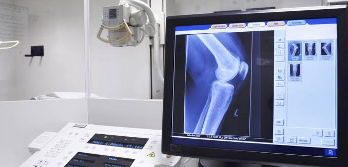
Here is how

01
Heavy-Duty, Hospital-Grade Components
Upright radiography demands durable mechanical hardware. Because patients must stand for weight-bearing pelvic, cervical, and lumbar views, your equipment faces constant physical adjustments throughout the day. Heavy-Duty Column Stands: Floor-to-wall or floor-to-ceiling tube tracks must deliver perfectly smooth, vibration-free movement to prevent motion artifacts during full-spine positioning. High-Frequency Generators: Upright full-spine imaging requires penetrative consistency through dense anatomical areas (like the lumbar-pelvic region) without extending exposure times. Advanced high-frequency generators minimize patient dose while capturing pristine cortical bone detail.
02
A Tailored Footprint for Private Practices
Unlike sprawling hospital radiology wings, private chiropractic clinics must carefully optimize their square footage. Compact Space Configurations: A dedicated chiropractic setup maximizes internal room dimensions. By pairing advanced generators with flexible single flat-panel DR configurations, clinics can successfully execute comprehensive cervical, thoracic, lumbar, and full-spine studies without sacrificing valuable floor space. CR and DR Versatility: Whether retrofitting an existing analog room with a high-capacity Computed Radiography (CR) reader or investing in a state-of-the-art Direct Radiography (DR) digital panel, the footprint should remain clean, unencumbered, and highly professional.
03
Pristine Contrast for Advanced Biomechanical Analysis
Accurately identifying structural subluxations, joint misalignments, spinal curvatures, or degenerative changes requires exceptional image contrast and custom post-processing tools. Advanced Digital Software: Modern digital software packages are explicitly optimized for musculoskeletal visualization. Practitioners can seamlessly zoom, fine-tune window levels, adjust exposure metrics dynamically, and map spinal listings instantly at the workstation workstation. Patient Education Visuals: Presenting patients with crystal-clear, annotated digital X-rays provides undeniable visual evidence of their baseline structural health and provides a clear benchmark to track adjustment progress over time.